What happens when mitochondria never rest. measuring the effects of continuous stimulation

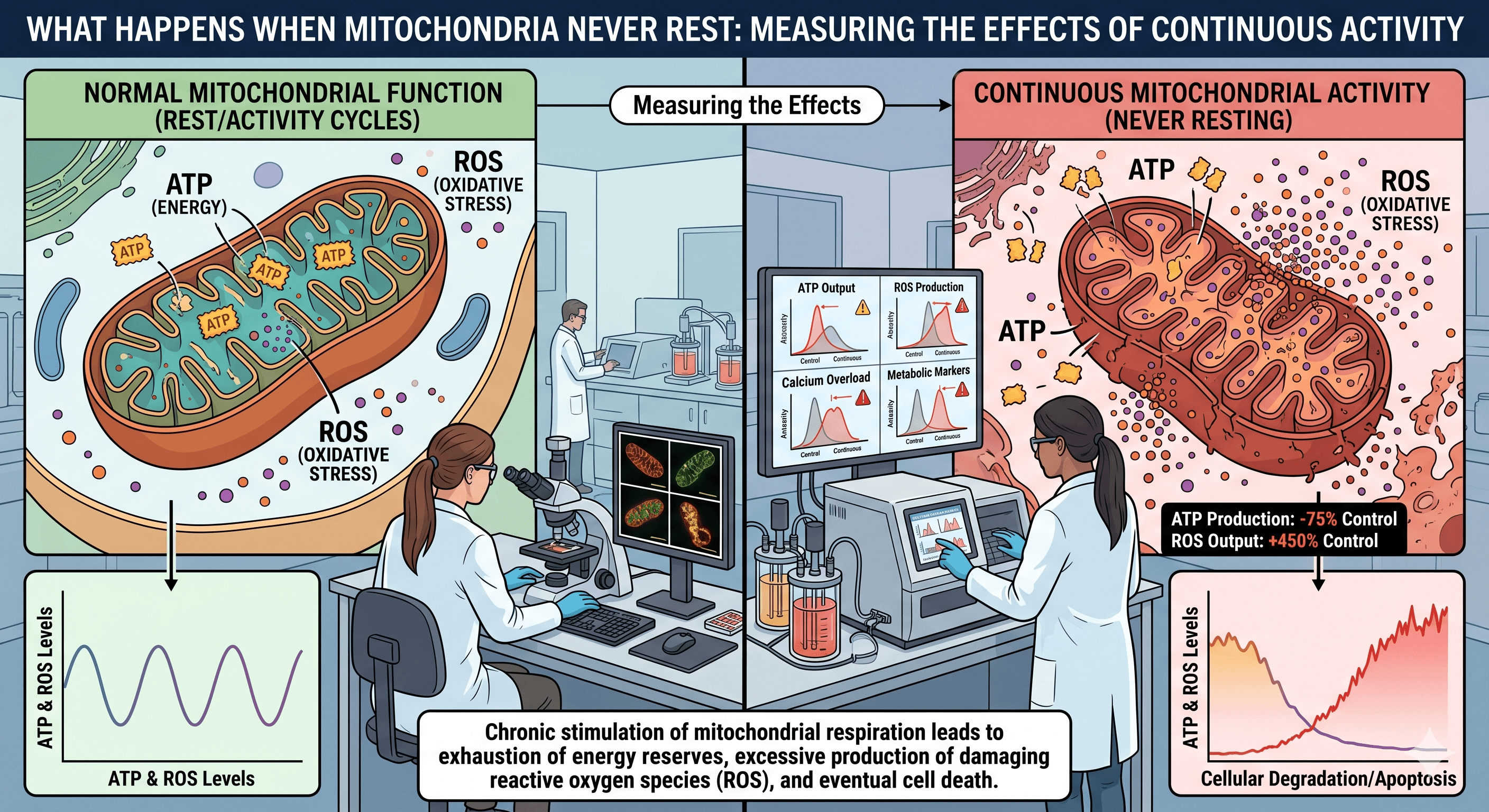
Mitochondria occupy a peculiar position in cell biology. They produce most of the ATP that powers eukaryotic life, yet they also generate reactive byproducts, regulate apoptosis, buffer calcium, and emit signals that reshape gene expression in the nucleus. Because of this dual role, the question of what happens when mitochondrial activity is continuously stimulated is not a simple bioenergetic puzzle. It is a question about how cells balance output against damage over time.
Defining continuous stimulation
In experimental biology, continuous stimulation of mitochondria can be achieved through several distinct interventions. Pharmacological uncouplers such as DNP or BAM15 dissipate the proton gradient, forcing the electron transport chain to run faster to maintain membrane potential. Substrate loading with pyruvate, succinate, or fatty acids increases flux through the tricarboxylic acid cycle. Optogenetic tools that depolarize the inner membrane on demand provide finer temporal control. Genetic approaches, such as overexpression of PGC-1α, push cells toward sustained biogenesis and elevated respiratory capacity.
Each of these methods stimulates a different aspect of mitochondrial function. Comparing them is essential, because results that hold for an uncoupler may not hold for a biogenesis driver. The phrase "stimulating mitochondria" is therefore an umbrella that conceals important mechanistic distinctions.
The short-term response
In the first minutes to hours after stimulation begins, cells typically show increased oxygen consumption, elevated ATP turnover, and a measurable rise in heat output. The NAD+/NADH ratio shifts, which in turn modulates the activity of sirtuins and other redox-sensitive enzymes. Calcium uptake into the matrix increases, sharpening the activity of dehydrogenases that depend on it.
If the stimulation is mild, the cell adapts gracefully. Antioxidant defenses including glutathione peroxidase, peroxiredoxins, and superoxide dismutase are upregulated. Mitochondrial dynamics shift toward fusion, which is thought to dilute damaged components across a larger network and protect respiratory efficiency. This phase resembles classical mitohormesis, where a modest stressor improves overall cellular fitness.
The long-term picture
Beyond a few days of unrelenting stimulation, the picture changes. Studies in cultured myotubes, primary hepatocytes, and whole organisms such as Caenorhabditis elegans and mice show a consistent pattern. Initial gains in oxidative capacity plateau, then decline. The decline correlates with several measurable changes:
Cumulative oxidative damage. Persistent electron flux raises the steady-state concentration of superoxide and hydrogen peroxide. Over time, this damages mitochondrial DNA, which lacks the protective histones of nuclear DNA and lies close to the source of reactive oxygen species. Mutation burden in mtDNA rises measurably under sustained high flux conditions.
Proteostatic strain. The mitochondrial unfolded protein response, mediated in mammals by ATF5 and in nematodes by ATFS-1, is activated early and remains active. Chronic activation of this pathway is itself associated with reduced lifespan in several models.
Membrane remodeling. Cardiolipin, the signature lipid of the inner membrane, becomes progressively oxidized. Loss of native cardiolipin disrupts the assembly of supercomplexes in the electron transport chain, reducing efficiency and increasing electron leak — a self-reinforcing loop.
Metabolic rewiring. Cells often shift toward glycolysis to spare overworked mitochondria, a phenomenon visible in increased lactate production even under normoxic conditions. Paradoxically, stimulation aimed at boosting oxidative metabolism can end in a more glycolytic phenotype.
What the measurements actually show
Researchers quantifying these effects rely on a converging set of tools. Seahorse extracellular flux analysis tracks oxygen consumption rate and extracellular acidification in real time. MitoSOX and genetically encoded sensors such as HyPer report on reactive oxygen species in living cells. Mass spectrometry quantifies oxidized proteins and lipids. Long-read sequencing of mtDNA detects deletions and point mutations that accumulate with chronic stress.
The picture that emerges across these measurements is not one of simple improvement or simple harm. It is dose-dependent and time-dependent. Brief or intermittent stimulation, particularly when interspersed with recovery periods, tends to produce durable benefits in respiratory capacity and stress resistance. Continuous, high-intensity stimulation tends to exhaust the adaptive machinery and accelerates the very dysfunctions it was intended to prevent.
Implications for intervention
The therapeutic interest in mitochondrial stimulation is substantial. Conditions ranging from sarcopenia and type 2 diabetes to neurodegeneration involve some component of mitochondrial decline. The data on continuous stimulation suggest that pulsatile or cyclic interventions are likely to outperform constant ones. Exercise, fasting, and cold exposure all impose intermittent mitochondrial stress followed by recovery, and all show consistent benefits across species.
The lesson is that the organelle thrives on rhythm, not relentless drive. Measurement, in this domain, is less about finding a maximum than about identifying the boundary where stimulation stops producing adaptation and begins producing damage.

Chamika Bandara
Author